All research

Research theme 2

Cell-free Synthetic Biology

We build high-yielding cell-free systems from diverse bacteria to make peptides, proteins and small molecules, and to prototype pathways quickly.

A cell-free system is a cell extract that carries out gene expression in a test tube. It holds the ribosomes, enzymes and metabolism of a living cell, but contains no living cell. That makes it fast, open to measurement and easy to automate.

We develop high-yielding extracts for academic and industrial use, and our best systems reach up to 4 mg/mL of peptide or protein. Our toolkit spans model organisms such as E. coli and non-model species including Streptomyces venezuelae and Klebsiella pneumoniae.

Liquid-handling robots let us test hundreds of conditions in one plate. We use cell-free reactions to screen genetic parts, build biosensors and prototype biosynthetic pathways without the constraints of living cells.

We co-organise the London Cell-free Biological Products Network. It meets twice a year at Imperial and UCL and brings together 30 to 50 researchers from academia and industry.

Key innovation: we are developing high-yielding, affordable cell-free systems for engineering peptides, proteins and small molecules through cell-free biosynthesis.
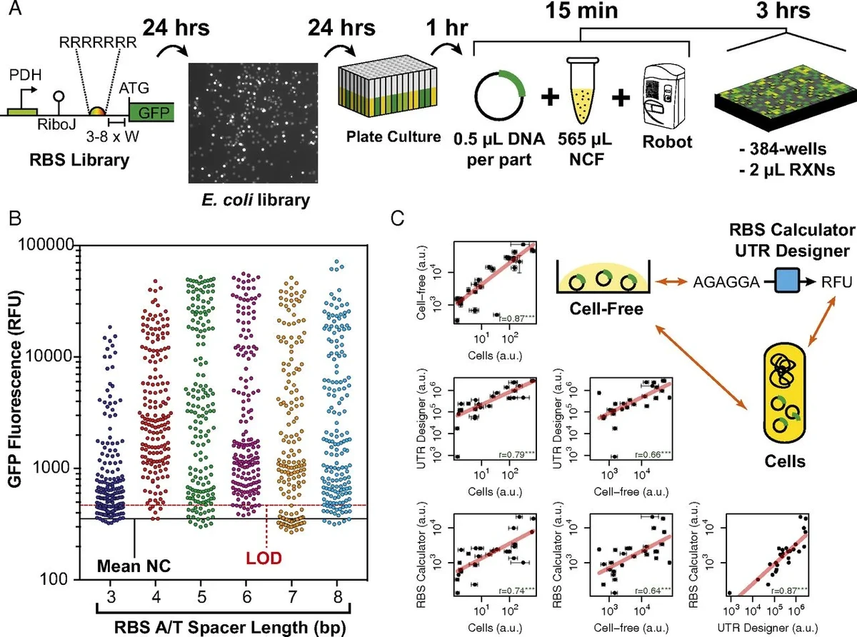
Figure showing a ribosome binding site library workflow, GFP fluorescence by spacer length, and comparisons of cell-free expression with predicted values.
Figure from Moore et al., PNAS (2018). doi:10.1073/pnas.1715806115 (opens in a new tab)

Key numbers

  • 4 mg/mLpeptide or protein from our best cell-free systems
  • 384wells per plate in our automated reactions, at 2 µL each
  • 266 µg/mLprotein from our Streptomyces venezuelae extract (ACS Synth. Biol. 2021)

How cell-free works

How we make and test a cell-free reaction

A cell-free system uses the machinery of a cell without the cell. First we make the extract, then we use it to make protein.
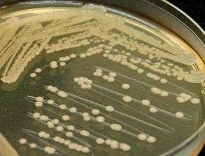
A. Making the extract

  1. Bacterial colonies growing on an agar plate.

    Grow

    We grow bacteria such as E. coli or Streptomyces venezuelae and harvest them at the growth phase that gives the most active extract.
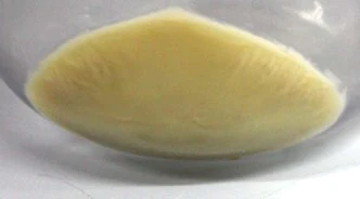
  2. A pale cream cell pellet lying in a dish after centrifugation.

    Harvest

    We collect the cells by centrifugation. The pellet is the starting material for the extract.
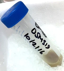
  3. A tube with a blue cap resting on ice, holding a small cell pellet and labelled in marker pen.

    Wash

    We wash the pellet and keep the tube cold, ready for lysis.
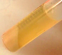
  4. A tilted tube holding clear amber cell extract.

    Lyse, spin and finish the extract

    We break open the cells, add a heat incubation step where the protocol needs one, and spin down the debris. The clear extract holds the ribosomes, enzymes and other machinery that a living cell uses to make protein.
B. Running and testing the reaction

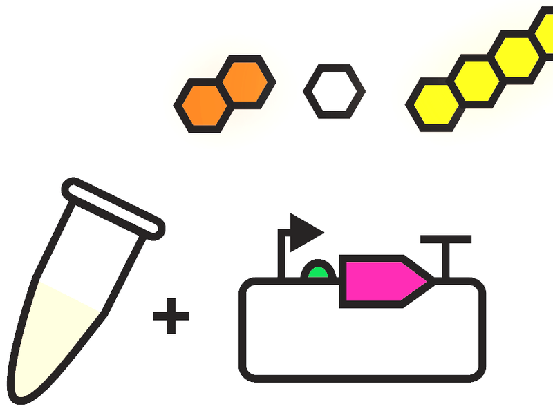
  1. Cartoon of an extract tube plus a plasmid carrying a pink gene, with orange, white and yellow hexagons for the substrates.

    Add DNA and substrates

    We add the DNA that encodes our protein or pathway, with the energy sources, amino acids and salts that fuel the reaction.
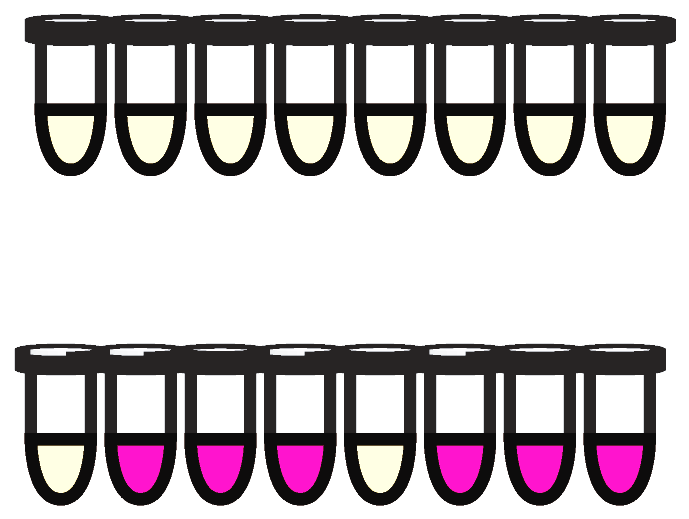
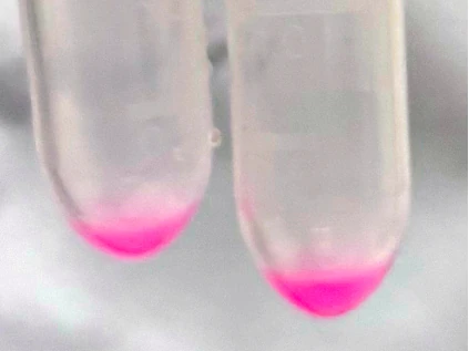
  2. Cartoon of two rows of eight tubes. All tubes in the top row are pale; in the bottom row six tubes are pink and two are pale.Two clear tubes, each with a bright pink layer at the bottom where the E. coli cell-free reaction has made mScarlet-I.

    Express

    The extract transcribes the DNA and translates the messenger RNA. When the gene encodes the fluorescent protein mScarlet-I, the reaction turns pink (right: our E. coli cell-free reactions).
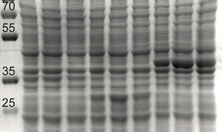
  3. Coomassie-stained protein gel of E. coli cell-free reactions, with a size marker at the left labelled 70, 55, 35 and 25. The three right-hand lanes show an intense band just above the 35 marker, and the middle lanes an extra band near 25, bands that the left-hand control lanes lack.

    Check the protein

    SDS-PAGE is used to analsye overproduced proteins. Reactions that make a protein show an enlarged band when compared to the control.
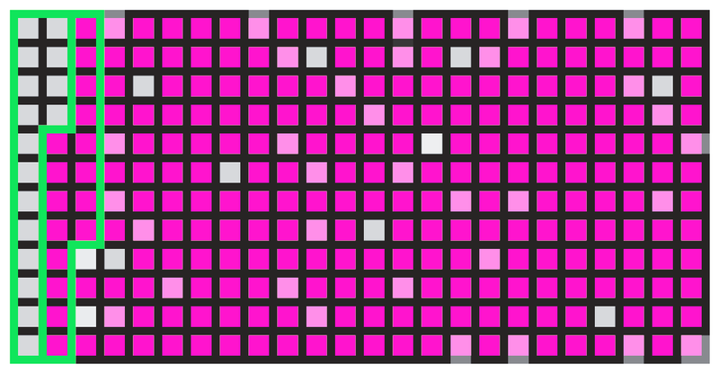
  4. Cartoon of a 384-well plate with most wells pink and a few pale.Cartoon of a plate reader.

    Read out and automate

    We read fluorescence or enzyme activity in a plate reader. Liquid-handling robots set up many conditions in parallel in 384-well plates.

Photographs and cartoons: Moore group. Gel: Nagappa et al., Front. Bioeng. Biotechnol. (2022) 10:992708 (opens in a new tab), CC BY.

Protein production

How much protein a cell-free reaction makes

Cell-free reactions make enough protein to see, measure and purify. These data come from reactions in the Moore group.
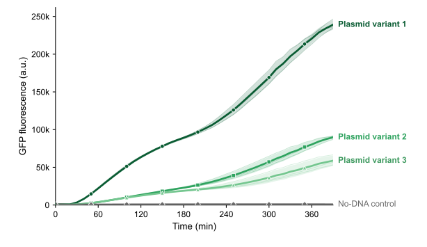
Line chart of GFP fluorescence against time over 390 minutes in E. coli cell-free reactions. Three plasmid variants rise steadily to about 239,000, 90,000 and 59,000 units, while the no-DNA control stays near 900.
GFP fluorescence over time. E. coli cell-free reactions with 10 nM plasmid DNA, showing continious activity for over 6 hours.
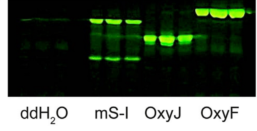
In-gel fluorescence of E. coli cell-free reactions. Bright green bands mark mScarlet-I, OxyJ and OxyF, in three lanes each, and the water control has almost none.
Protein bands in the gel. E. coli Rosetta 2 (DE3) reactions make C-terminally tagged mScarlet-I, and biosynthetic enzymes, which an in-gel fluorescent stain. The control (ddH2O) shows no bands. This experiment showed that tryptone, yeast extract and casamino acids are suitable low cost replacements for pure amino acids in cell-free protein synthesis. Adapted from Nagappa et al., Front. Bioeng. Biotechnol. (2022) (opens in a new tab), CC BY.
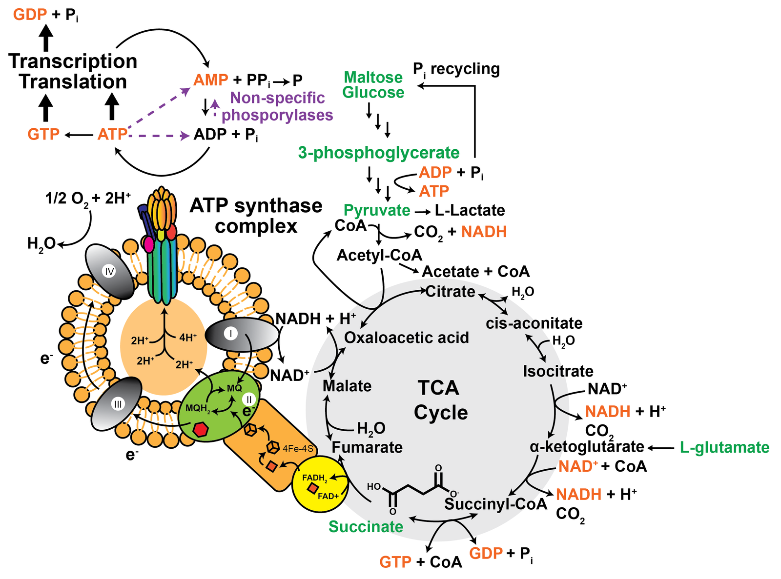
Diagram of energy and metabolism in an E. coli cell-free reaction. Transcription and translation consume ATP and GTP, which the extract regenerates from sugars, 3-phosphoglycerate and glutamate through glycolysis, the TCA cycle and membrane vesicles with an ATP synthase complex.
How the reaction is fuelled. Transcription and translation require ATP and GTP. An E. coli cell extract regenerates the energy molecules from added substrates (green) through glycolysis, the TCA cycle and inverted membrane vesicles that have an ATP synthase complex. Figure © Simon J. Moore, from Rice et al., Chem. Soc. Rev. (2025) 54:4314-4352 (opens in a new tab).

Automation

Robots let us test hundreds of reactions

Cell-free reactions can be automated using liquid-handling robots. We have published different automated approaches to assemble 384-well plates with 2-10 µL reactions and measured ribosome binding site variants and promoters from Bacillus megaterium (Moore et al., PNAS 2018), as well as antimicrobial resitance (Bergum et a., Synthetic Biology 2026).

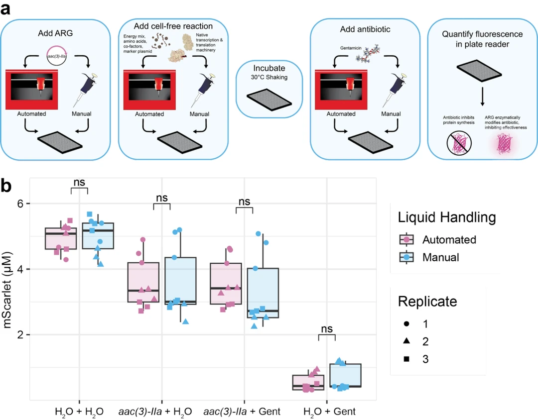
We use a small Myra robotic system in preference to manual pipetting for preparing 384 plate reactions. We compared human versus robot and found no statistical difference. We used the workflow to show that antibiotic resistance genes change the potency of antibiotics, which links this theme to our antimicrobial resistance work (Bergum et a., Synthetic Biology 2026).

Read the papers: Bergum et al., Synthetic Biology (2026) 11:ysag010 (opens in a new tab), and the Methods in Enzymology chapter (2026, doi:10.1016/bs.mie.2026.09.046 (opens in a new tab)).

Selected publications

Each entry links to the paper, its Altmetric record and a live citation count.

  • Publication 30

    Molly Bergum, Sahana Suthakaran, Bethany Martin, J. Mark Sutton, Simon J. Moore

    A liquid handling platform for standardized quantification of cell-free enzymatic activity encoded by antimicrobial resistance genes

    Synth. Biol. (2026) 11:ysag010

    Workflow diagram and box plot. The diagram shows five steps in rounded boxes: add an antimicrobial resistance gene plasmid, add the cell-free reaction, incubate at 30 degrees Celsius with shaking, add antibiotic, and quantify fluorescence in a plate reader, with an automated liquid handling robot and a manual pipette shown as alternatives for each addition. Below it, a box plot of mScarlet concentration for four reaction mixtures shows no significant difference between automated and manual pipetting.
  • Publication 28

    Andrew J. Rice, Tien T. Sword, Kameshwari Chengan, Douglas A. Mitchell, Nigel J. Mouncey, Simon J. Moore and Constance B. Bailey

    Cell-free synthetic biology for natural product biosynthesis and discovery

    Chemical Society Reviews (2025) 54:4314-4352
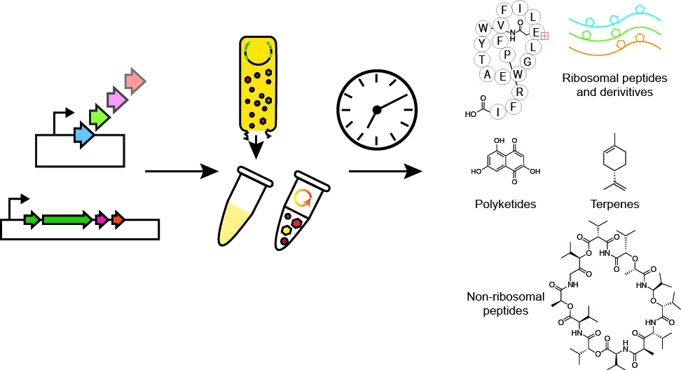
    Schematic of cell-free biosynthesis: DNA constructs and extract in tubes lead to natural product classes including ribosomal peptides, polyketides, terpenes and non-ribosomal peptides.
  • Publication 23

    Lakshmeesha K. Nagappa, Wakana Sato, Farzana Alam, Kameshwari Chengan, Christopher M. Smales, Tobias Von Der Haar, Karen M. Polizzi, Katarzyna P. Adamala and Simon J. Moore

    A ubiquitous amino acid source for prokaryotic and eukaryotic cell-free transcription-translation systems

    Front. Bioeng. Biotechnol. (2022)10:992708
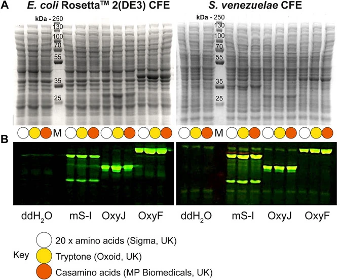
    Gels and fluorescence read-outs comparing cell-free extracts from E. coli Rosetta 2(DE3) and Streptomyces venezuelae with different amino acid sources.
  • Publication 22

    Agata Kennedy, Guy Griffin, Paul S. Freemont, Karen M. Polizzi, and Simon J. Moore

    A curcumin direct protein (DiPro) biosensor for cell-free prototyping

    Engineering Biology 6(2):62-68 (2022)
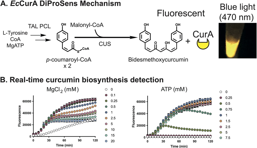
    Curcumin biosensor: the biosynthesis pathway, a fluorescent signal under blue light, and real-time fluorescence curves at different magnesium and ATP concentrations.
  • Publication 16

    Simon J. Moore, Hung-En Lai, Soo-Mei Chee, Ming Toh, Seth Coode, Kameshwari Chengan, Patrick Capel, Christophe Corre, Emmanuel de Los Santos, and Paul S. Freemont

    A Streptomyces venezuelae Cell-Free Toolkit for Synthetic Biology

    ACS Synth. Biol. (2021) 10: 402-411

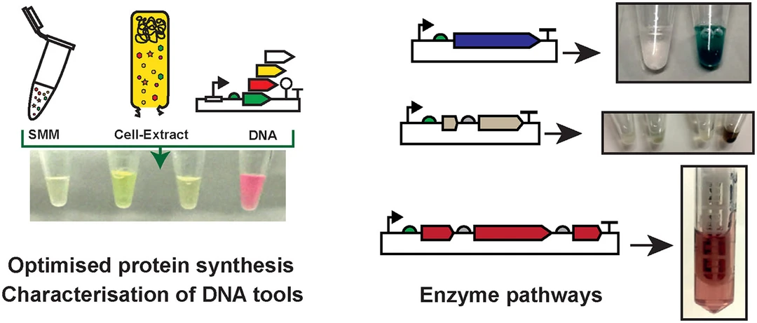
    Streptomyces venezuelae cell-free toolkit: optimised protein synthesis, characterisation of DNA tools, and enzyme pathway reactions that give coloured products.
  • Publication 13

    Simon J. Moore, James T. MacDonald, Sarah Wienecke, Alka Ishwarbhai, Argyro Tsipa, Rochelle Aw, Nicolas Kylilis, David J. Bell, David W. McClymont, Kirsten Jensen, Karen M. Polizzi, Rebekka Biedendieck, and Paul S. Freemont

    Rapid acquisition and model-based analysis of cell-free transcription-translation reactions from non-model bacteria

    Proceedings of the National Academy of Sciences, USA (2018) 115:E4340-E4349
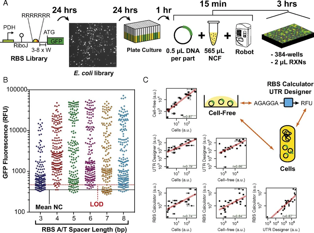
    Ribosome binding site library workflow, GFP fluorescence by spacer length, and comparisons of cell-free expression with predicted values.
More from this theme (8)
  • Publication 24

    Simon J. Moore, Hung-En Lai, Jian Li and Paul S. Freemont

    Streptomyces cell-free systems for natural product discovery and engineering

    Nat. Prod. Rep. (2023) 40:228-236

  • Publication 21

    Jian Li, Yong-Chan Kwon, Yuan Lu, Simon J. Moore

    Editorial: Cell-Free Synthetic Biology

    Frontiers in Bioengineering and Biotechnology 9:799122 (2021)

  • Publication 19

    Ming Toh, Kameshwari Chengan, Tanith Hanson, Paul S. Freemont, and Simon J. Moore

    A high-yield Streptomyces transcription-translation toolkit for synthetic biology and natural product applications

    JoVE (2021) 10.3791/63012

  • Publication 18

    Simon J. Moore, Tomasso Tosi, David J. Bell, Yonek Hleba, Karen M. Polizzi, and Paul S Freemont

    High-yield 'one-pot' biosynthesis of raspberry ketone, a high-value fine chemical

    Synth. Biol. (2021) 6:ysab021

  • Publication 15

    Simon J. Moore

    Enzyme alchemy: cell-free synthetic biochemistry for natural products

    Emerging Topics in Biosciences (2019) 3: 529-535

  • Publication 12

    Simon J. Moore, James T. MacDonald, and Paul S. Freemont

    Cell-free synthetic biology for in vitro prototype engineering

    Biochemical Society Transactions (2017) 45:785-791

  • Publication 10

    Simon J. Moore, Hung-En Lai, Hannah Needham, Karen M. Polizzi, and Paul S. Freemont

    Streptomyces venezuelae TX-TL - a next generation cell-free synthetic biology tool

    Biotechnol J (2017) 12:1600678

  • Publication 3

    Molly Bergum, Bethany Martin, J. Mark Sutton, Simon J. Moore

    A semi-automated cell-free protein synthesis workflow for quantifying antibiotic-inactivating enzymes

    Methods in Enzymology (2026) DOI: 10.1016/bs.mie.2026.09.046

All publications

More research themes

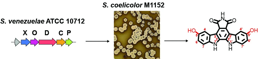
Figure from the arcyriaflavin F paper: the biosynthetic gene cluster in Streptomyces venezuelae, a plate of Streptomyces coelicolor M1152 colonies, and the structure of arcyriaflavin F.

Theme 1

Synthetic Biology for Natural Products

We engineer Streptomyces bacteria to discover and make new bioactive molecules, from single enzymes to complete pathways.
Work with us

We welcome enquiries from prospective students and fellows, and from industry and public-sector partners.

Join usContact details